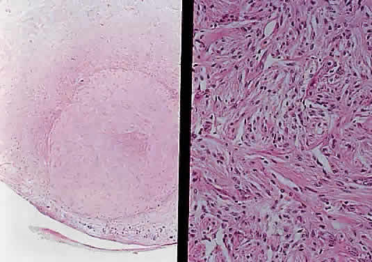
Fig. 37.
Pilocytic glioma of optic nerve (hematoxylin and eosin staining). Low power on left shows nerve surrounded by thickened meninges. High power on right shows spindle-shaped cells.