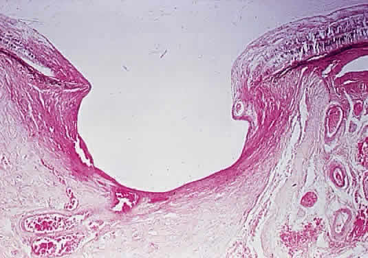
Fig. 32.
Deeply cupped optic disc. Deep posterior bowing of laminar cribrosa and loss of tissue anterior to lamina (hematoxylin and eosin staining)