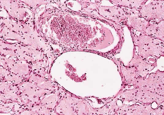
Fig. 9.
Thick-walled central retinal artery and vein in common thin pial sheath (cross-section hematoxylin and eosin staining).