|
|
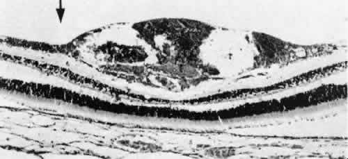
| Fig. 6. Intraretinal submembranous hemorrhage shows blood between the retinal internal limiting membrane and the nerve fiber layer. Arrow indicates the surface of the posteriorly detached vitreous (posterior hyaloid) (hematoxylin and eosin, original magnification, × 70) |