|
|
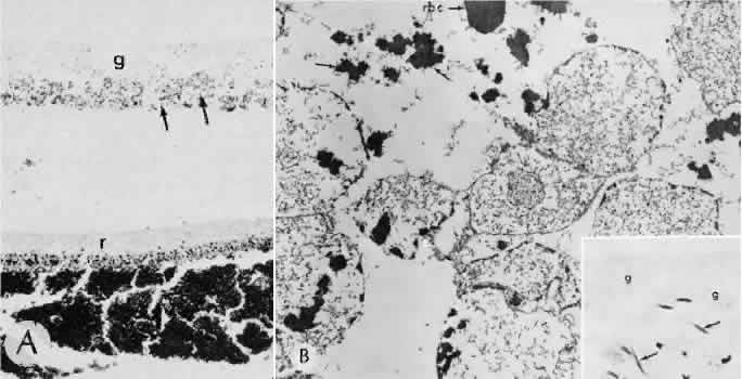
| Fig. 4. Hemorrhage into the eye. A. Erythrocytes beneath the retina ® appear normal; those in the vitreous (arrows) are grossly sickled. The deepest part of the vitreous is occupied by erythrocyte ghosts (g) because of the intense hemolysis in this relatively hypoxic region (see B). B (Inset). Grossly sickled erythrocytes (arrows) in the vitreous. The deeper region contains hemolyzed erythrocytes (ghosts). Electron micrograph shows almost complete hemolysis of the erythrocytes (ghosts), free clumps of hemoglobin outside the cells (arrows), and the tip of a sickled, nonhemolyzed cell (rbc) nearby. (A, hematoxylin and eosin, × 85; B, original magnification, × 15,000; inset, hematoxylin × 530) |