|
|
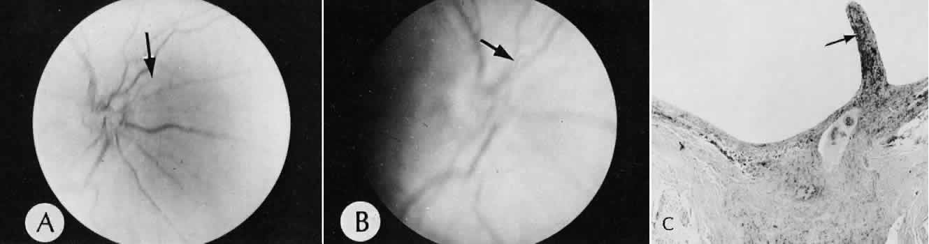
| Fig. 1. Posterior remnants. A. Fundus photograph with the focus just anterior to the optic disc to show a persistent hyaloid vessel (arrow) in a 22-year-old man. B. Focus in the posterior part of the vitreous to show the hyaloid vessel (arrow) running anteriorly, where it inserts on the posterior aspect of the lens C. Bergmeister's papilla, a glial remnant of the hyaloid system, persists (arrow) in an adult eye. (A, fundus; B, fundus; C, hematoxylin and eosin, original magnification, × 16) |