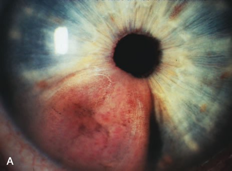
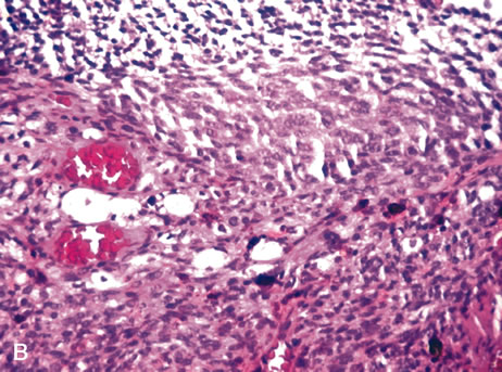
Fig. 4.
Iris melanoma. A pigmented mass involves the iris (A), which histologically reveals spindle cells (B). (Hemotoxylin-eosin ×60.)