|
|
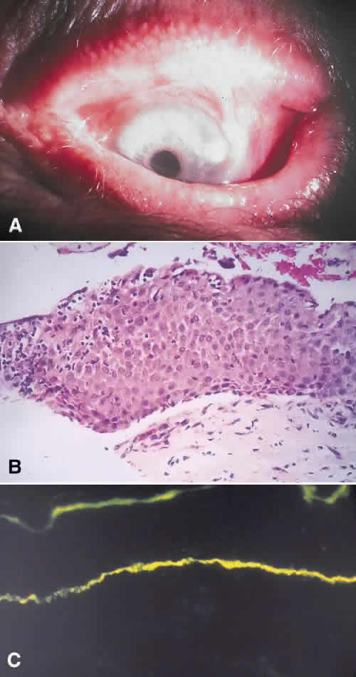
| Fig. 10. A. Clinical photograph shows cicatricial changes in the bulbar and palpebral conjunctiva with symblepharon in benign mucosal pemphigoid. B. Histopathology shows epithelial bullae with a slight perivascular, lymphocytic infiltrate in the substantia propria. C. Immunofluorescent stains show linear deposition of IgG and IgA along the basement membrane zone of the conjunctival epithelium. |