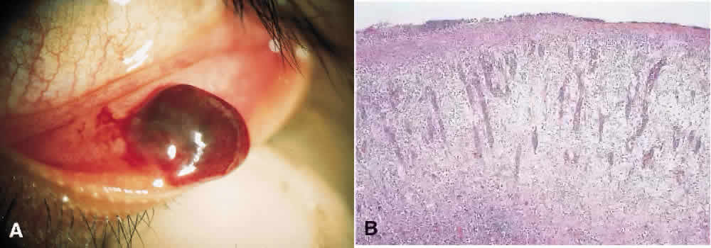
Fig. 8.
A.
Clinical photograph of nodular, fleshy, pedunculated, red mass located in the inferior fornix.
B.
Histopathology shows radiating pattern of abundant capillaries and fibroblasts amid an edematous stroma.