|
|
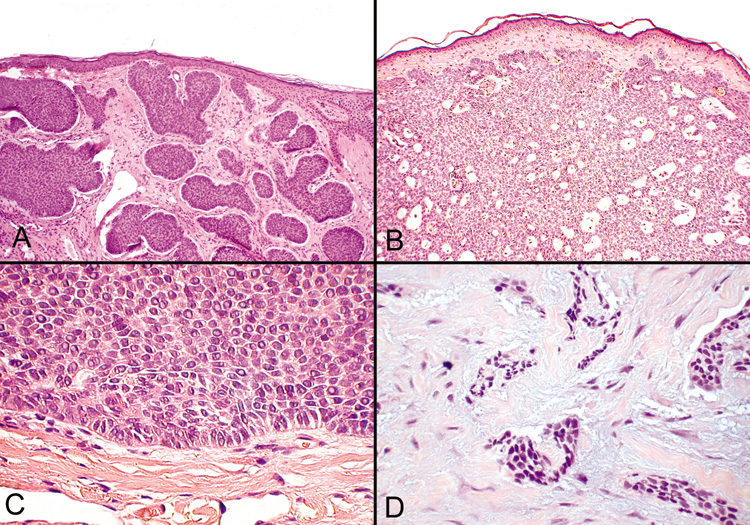
| Fig. 45. Basal Cell Carcinoma—A, B. Photomicrographs of nodular basal cell carcinomas. C. Typical palisading of tumor cells along the periphery of the tumor nodule. D. The morpheaform variant shows longs strands or clumps of tumor cells invading a dense fibrous stroma (hematoxylin and eosin stain). (Photos courtesy of William Morris, M.D.) |