|
|
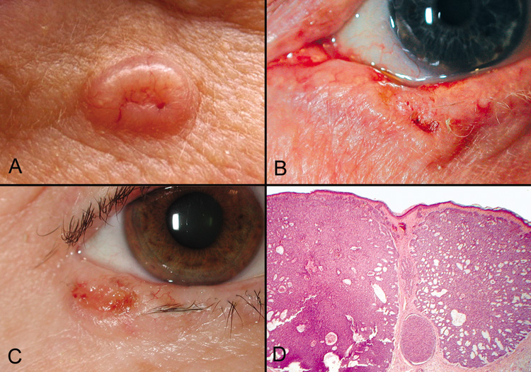
| Fig. 44. Basal Cell Carcinoma—A, B, C. Clinical photographs of various appearances of basal cell carcinomas, the typical “rodent ulcer.” Notice the loss of lashes at the tumor site and in adjacent areas. D. Low-power photomicrograph of nodular variant of basal cell carcinoma (hematoxylin and eosin stain). (Photos courtesy of William Morris, M.D.) |