|
|
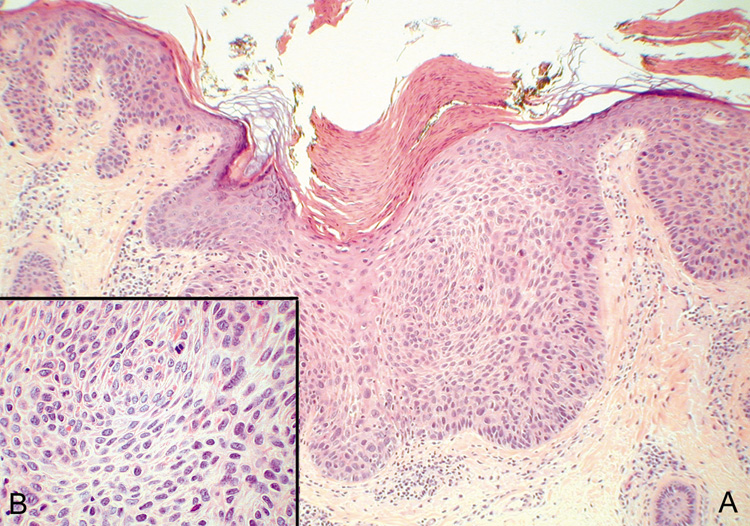
| Fig. 43. Carcinoma In Situ—A. Low-power photomicrograph demonstrating parakeratosis, hyperkeratosis, and epithelial dysplasia confined to the epithelium. No invasion is present. B. High-power photomicrograph revealing pleomorphic cells and active mitosis (hematoxylin and eosin stain). (Photos courtesy of William Morris, M.D.) |