|
|
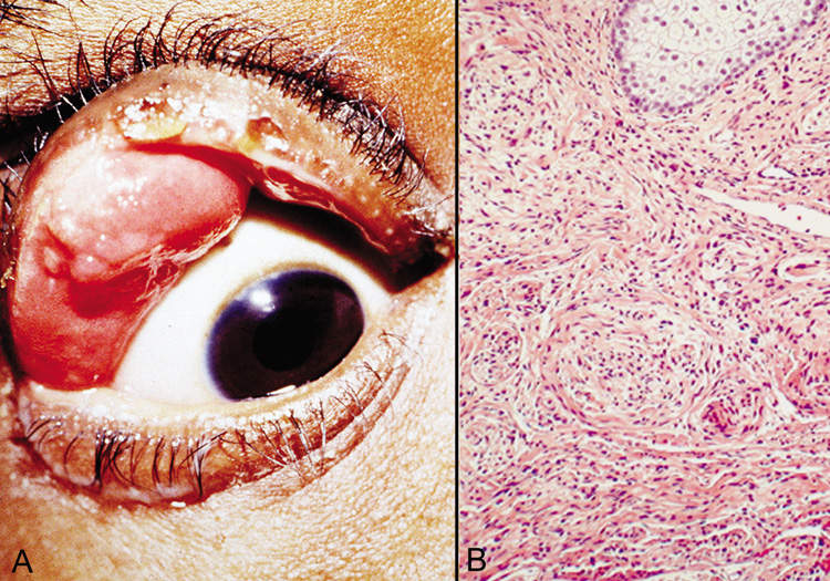
| Fig. 40. Plexiform neurofibroma—A. Clinical photograph of patient with plexiform neurofibroma involving the upper lid. B. Low-power photomicrograph showing enlarged abnormal nerves composed of endoneural fibroblasts, Schwann cells, and axons (hematoxylin and eosin stain). (Photos courtesy of William Morris, M.D.) |