|
|
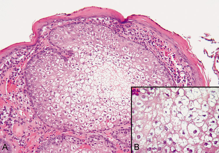
| Fig. 32. Sebaceous Adenoma—A. Low-power photomicrograph showing sebaceous nodule containing undifferentiated sebaceous cells in the periphery and mature sebaceous cells in the center (hematoxylin and eosin stain). B. High-power view of the central mature sebaceous cells (hematoxylin and eosin stain). (Photos courtesy of William Morris, M.D.) |