|
|
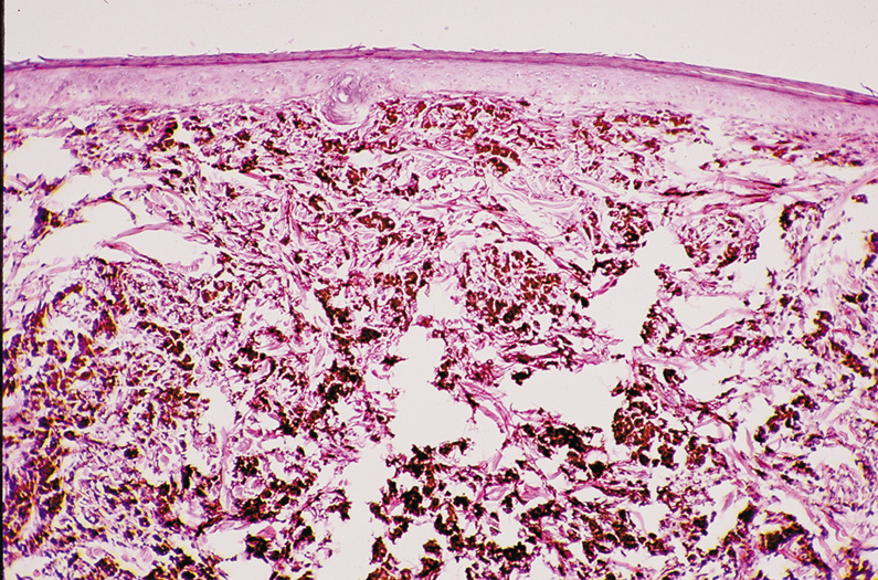
| Fig. 29. Dermal (Intradermal) Nevus—A, C. Clinical photographs of two different dermal nevi. A. Smooth, dome-shaped appearance. C. Papillary appearance. B. Proliferation of nests of nevus cells in dermis of skin with the larger more heavily pigmented cells more superficial than the smaller less pigmented nevus cells. No nevus cells are present within the overlying epithelium (hematoxylin and eosin stain). (Photos courtesy of William Morris, M.D.) |