|
|
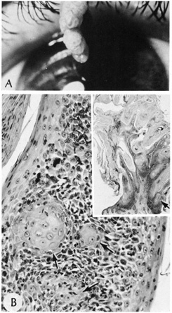
| Fig. 26. Inverted follicular keratosis. A. Clinical appearance. B. Inset shows hyperkeratotic papillomatous lesion shown under high magnification, (arrow main figure). Note: acantholytic squamous cells surrounding squamous eddies (arrows). (Modified from Sassani JW, Yanoff M: Inverted follicular keratosis. Am J Ophthalmol 87:810, 1979; and Scheie HG, Yanoff M, Sassani JW: Inverted follicular keratosis mimicking malignant melanoma. Ann Ophthalmol 9:949, 1977.) |