|
|
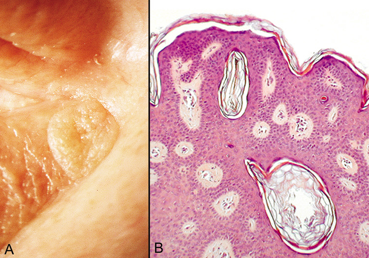
| Fig. 25. Seborrheic Keratosis—A. Clinical photograph of seborrheic keratosis illustrating its “stuck on“ appearance. B. Low-power photomicrograph showing the proliferating cords of basal cells, the vascular islands, and the “horn cysts“ within the thickened epithelium (hematoxylin and eosin stain). (Photos courtesy of William Morris, M.D.) |