|
|
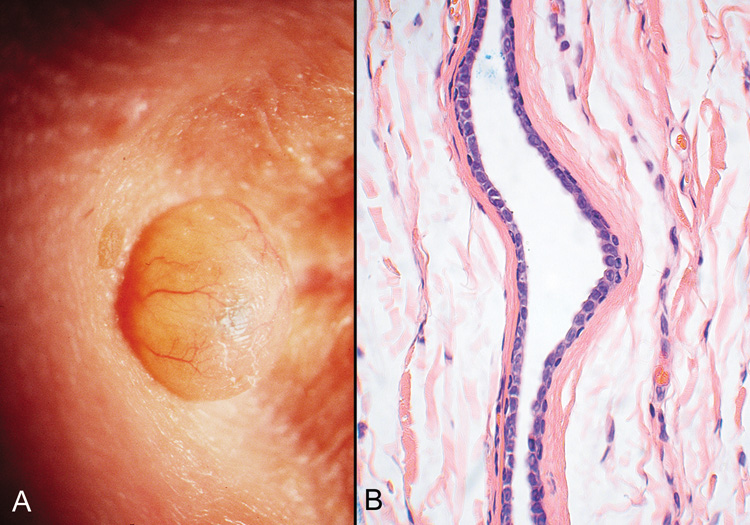
| Fig. 20. Eccrine Hidrocystoma—A. Clinically, this appears as a softly cystic lesion that transilluminates brightly. B. Photomicrograph showing collapsed cyst lined by a double row of cuboidal cells. The lumen appears empty (hematoxylin and eosin stain). (Photos courtesy of William Morris, M.D.) |