|
|
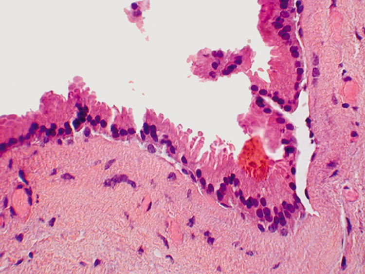
| Fig. 19. Apocrine Hidrocystoma—Apocrine cells line the hidrocystoma wall. Notice the “decapitation snouts“ at the apex of the cells, indicating that the cytoplasm has been pinched off to form the glandular secretion (hematoxylin and eosin stain). (Photo courtesy of William Morris, M.D.) |