|
|
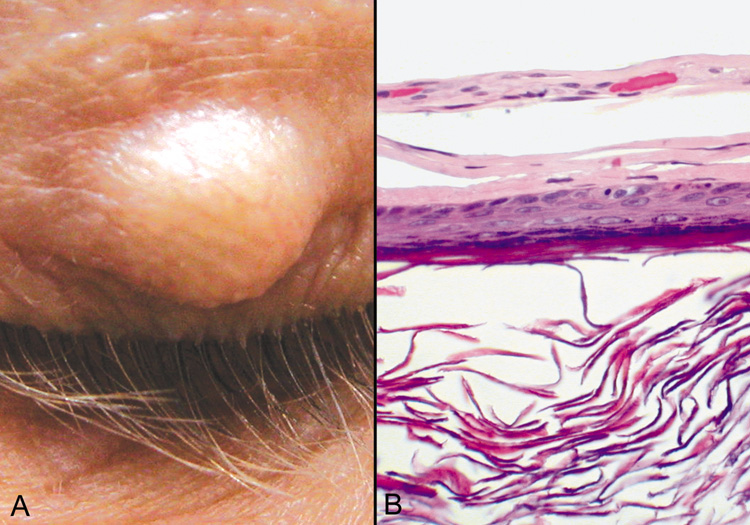
| Fig. 18. Epidermal Inclusion Cyst—A. Clinically this cystic lesion usually has a smooth dome-shape and is light yellow to white. B. Histopathologically, this cystic lesion is lined with stratified squamous epithelium that includes the granular cell layer. The lumen is filled with keratin produced by the epithelium (hematoxylin and eosin stain). (Photos courtesy of William Morris, M.D.) |