|
|
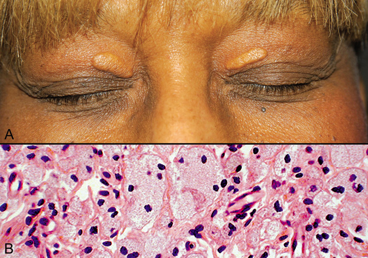
| Fig. 12. Xanthelasma—A. Clinical photograph of xanthelasma showing typical distribution of the xanthomatous nodules on the eyelids. B. High-power photomicrograph of many multinucleated foamy xanthoma cells (hematoxylin and eosin stain). (Photos courtesy of William Morris, M.D.) |