|
|
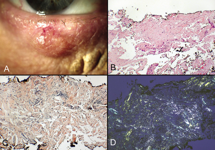
| Fig. 11. Amyloidosis—A. Erythematous appearance of amyloidosis involving the lid. This was incorrectly diagnosed as a chalazion. B. Amorphous eosinophilic material (amyloid) present within the excised tissue (hematoxylin and eosin stain). C. Positive staining of amyloid material with Congo Red. (Congo Red stain). D. When viewed with polarized light, apple-green birefringence of the Congo Red-stained amyloid deposits is visible (Congo Red stain/polarized light). (Photos courtesy of William Morris, M.D.) |