|
|
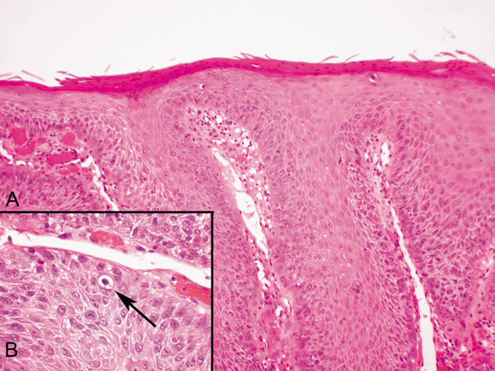
| Fig. 8. Verruca Vulgaris—A. Low-power photomicrograph illustrating papillomatous growth with a fibrovascular core, hyperkeratosis, and acanthosis (hematoxylin and eosin stain). B. High-power photomicrograph demonstrating intranuclear viral inclusion (black arrow) (hematoxylin and eosin stain). (Photos courtesy of William Morris, M.D.) |