|
|
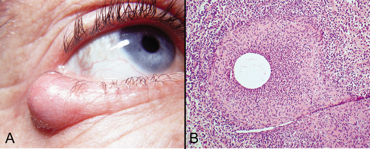
| Fig. 5. Chalazion—A. Typical clinical appearance of chalazion. B. Lipogranulomatous reaction with epithelioid cells, lymphocytes, and plasma cells surrounding a central nidus of Meibomian gland secretion. Clear area in center represents lipid material dissolved out during processing of tissue. Giant cells are sometimes seen (hematoxylin and eosin stain). (Photos courtesy of William Morris, M.D.) |