|
|
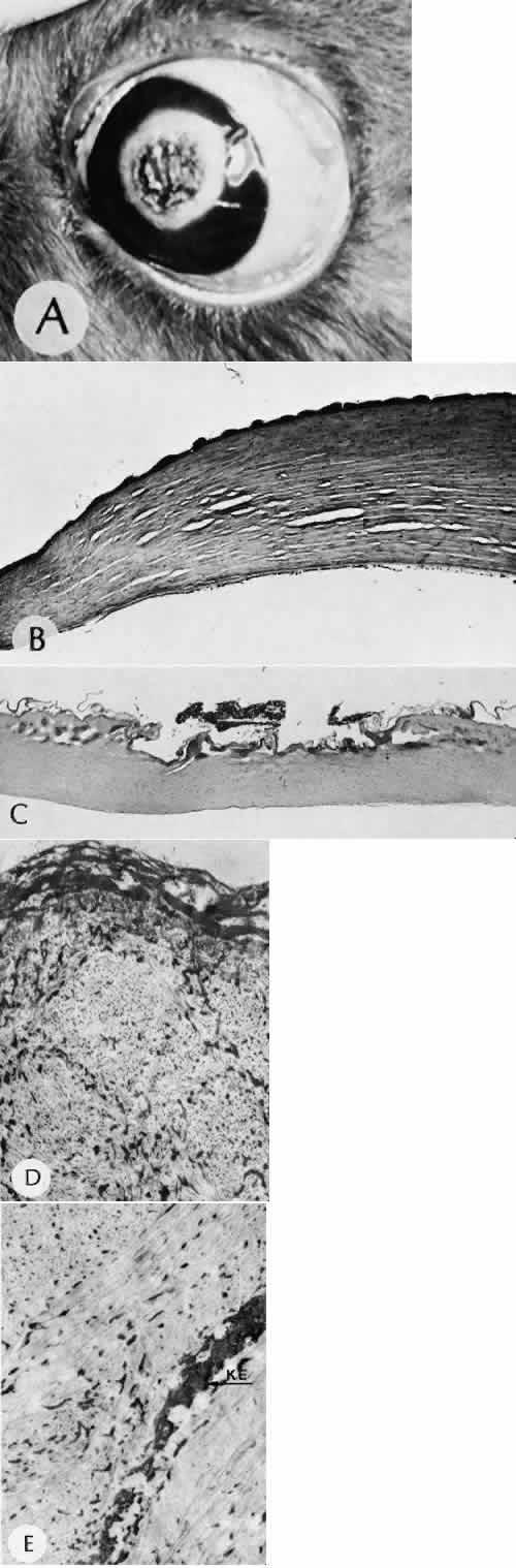
| Fig. 92. Carbon dioxide laser. A. Irradiated (35 w/cm2) rabbit eye showing charring of crater bed and white ring of adjacent cornea. B. Light micrograph shows three regions of corneal stroma: (1) relatively normal on extreme left, (2) edematous in middle (with clefts), and (3) grossly thickened with fused lamellae on right. The last accounts for intense white ring seen clinically. C. Central region of charred tissue in corneal burn (35 w/cm2). D. The portion of epithelium that underwent coagulative necrosis remains on surface. Subepithelial collagen shows widespread spotty densification (denaturation). E. Deep stroma shows spotty densification of collagen and coagulative necrosis of keratocyte (KE). (Hematoxylin-eosin stain; B, × 50; C, × 35; D and E, × 18,000 [25 w/cm2]. (B from Fine BS et al: Am J Ophthalmol 64:209, 1967.) |