|
|
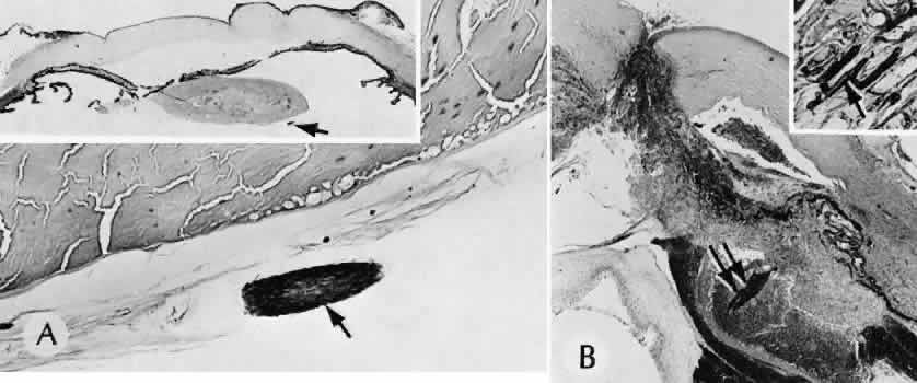
| Fig. 84. Intraocular foreign material. A. A cilium was implanted during penetrating trauma to the eye. The cilium can be seen behind a cataractous lens (arrow). (Hematoxylin-eosin stain; × 69) Inset. Retained cilium, posterior synechiae, iris bombe, and peripheral synechiae. (Hematoxylin-eosin stain; × 5.) B. A wooden foreign body is present in an abscess overlying the ciliary body in a second case of penetrating trauma to the eye. A ring of hemorrhage surrounds the abscess. (Hematoxylin-eosin stain; × 12.) Inset. The hyphae of a pigmented fungus (arrow) lie within the wood as an unsuspected potential source of infection. (Prussian blue stain; × 300.) (B from Fine BS. Lab Invest 11:1161, 1962.) |