|
|
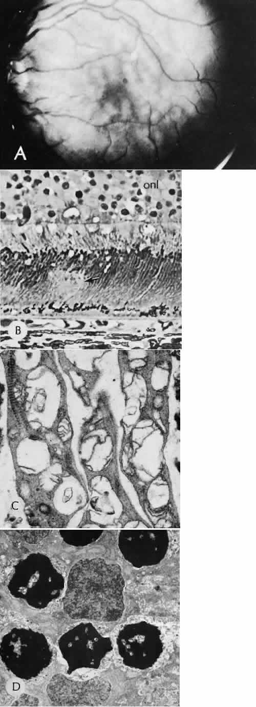
| Fig. 77. Commotio retinae. A. Throughout the posterior pole, patch areas of deep retinal opacification are present. The retina is attached, and the vascular system is competent. B—D, Transmission electron micrographs. B. The deep opacification corresponds with vacuolization of the inner portion of the photoreceptor layer (arrow). Many pyknotic nuclei are present in the outer nuclear layer (onl) in a person who died 48 hours after trauma. C. There is marked disruption of the mitochondria of the inner photoreceptors in this case 21 hours after ocular trauma. D. Higher magnification of the pyknotic nuclei, which are characterized by dense chromatic and an undulating nuclear membrane. (B, × 160; C, × 16,250; D, × 4050.) (Sipperley JO, Quigley HA, Gass DM: Traumatic retinopathy in primates: The explanation of commotio retinae. Arch Ophthalmol 96:2267–2273, 1978.) |