|
|
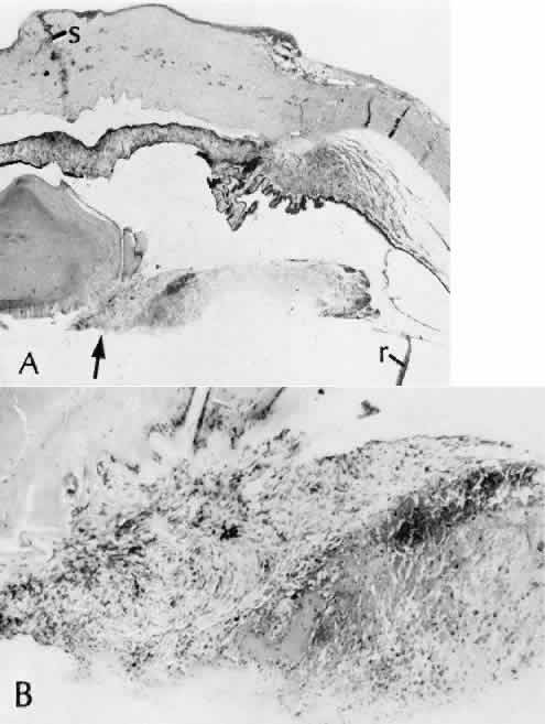
| Fig. 75. Cyclic membrane. In cases of extensive intraocular inflammation in the posterior chamber, the anterior vitreous face may act as a scaffolding over which fibrous tissue may proliferate and then contract. A. A corneal scar (s) of a perforating corneal wound is present in this histologic example. The injury produced hemorrhage and inflammation in the posterior chamber. The organization of the hemorrhage (arrow) is along the plane of the anterior vitreous face. Contraction of the fibrous tissue has already caused partial traction retinal detachment ®. B. Higher magnification of the region of the arrow in A shows the degree of fibroblastic proliferation. These cells have the ability to contract. The aggregate effect is to displace the neurosensory retina (traction retinal detachment). (Hematoxylin-eosin stain; A, × 11; B, × 28) |