|
|
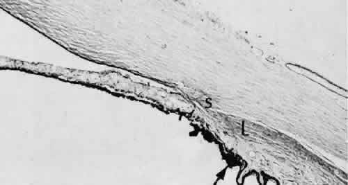
| Fig. 70. Angle recession with peripheral anterior synechiae. The characteristic fusiform shape of the normally wedge-shaped ciliary body is caused by atrophy of the round and oblique muscles of the ciliary body, leaving only the longitudinal (L) muscles intact. Iris neovascularization (rubeosis iridis) developed and caused secondary anterior peripheral synechia. The iris root (arrow) and anterior ciliary processes are displaced anteriorly from the scleral spurs. (Periodic acid-Schiff stain; × 16.) |