|
|
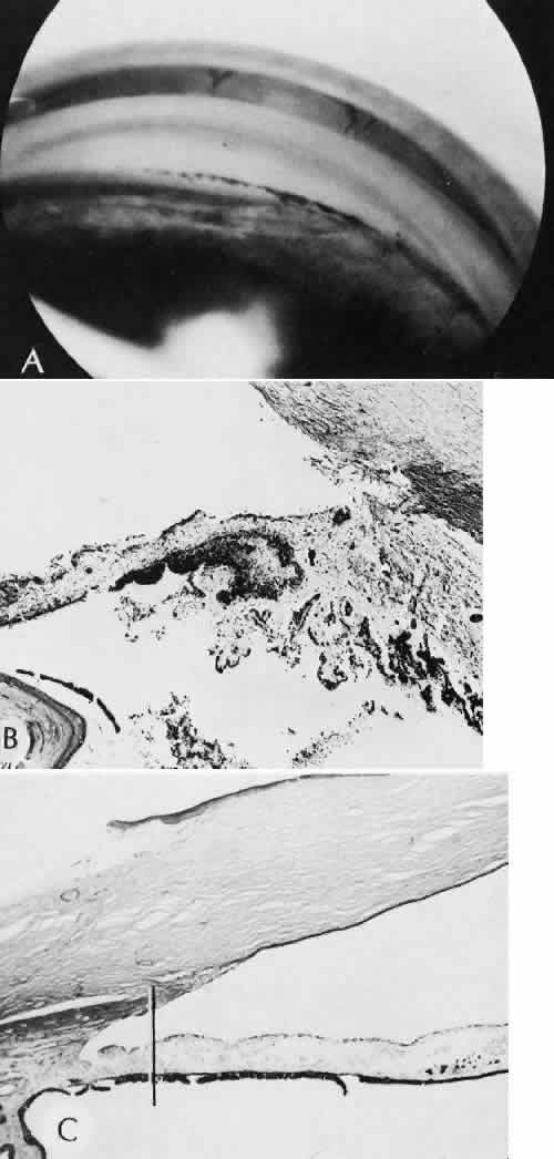
| Fig. 68. Contusion angle deformity. The plane between the longitudinal muscle of the ciliary body and the circular and oblique fibers of the ciliary body is another site of potential rupture when exposed to increased hydrostatic pressure within the anterior chamber. A. The characteristic gonioscopic appearance of contusion angle deformity is focal deepening of the anterior chamber angle. B. A laceration has occurred into the anterior face of the ciliary body. Blood is present in the anterior chamber and the supraciliary space. The longitudinal muscles of the ciliary body are still attached to the scleral spur. C. Fibrous contraction of scarred tissue results in posterior displacement of the iris root and pars plicata of the ciliary body. A line has been drawn through the scleral spur parallel to the optic axis. (Periodic acid-Schiff stain; × 16.) |