|
|
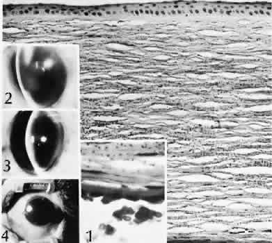
| Fig. 66. Blood staining of the cornea. Extravasated blood in the anterior chamber (hyphema) degenerates and liberates hemoglobin molecules. The hemoglobin migrates across an intact Descemet's membrane and diffuses into the corneal stroma. When an appropriate concentration is reached, the hemoglobin precipitates and forms multiple small particles throughout the area of diffusion. In this case, hemoglobin particles can be identified at all levels of the corneal stroma. (Brenner and Bren stain; × 101.) Inset 1. The size of the red blood cells and the stromal particles can be compared. (Brenner and Bren stain; × 441.) Insets 2–4. Various degrees of hyphema. If there is a sufficient volume of clotted blood, aqueous circulation is affected. Because of the lack of oxygen, the clot will turn from red to black (“black-ball” hyphema). |