|
|
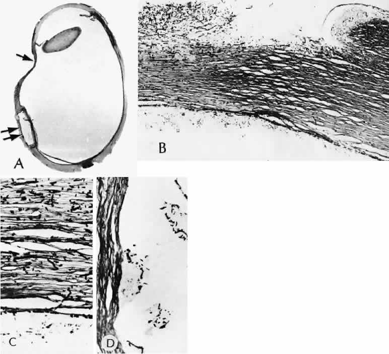
| Fig. 58. A case of fungal endophthalmitis following retinal reattachment surgery. A. Scleral thinning developed 3 weeks after retinal reattachment surgery (arrow). (Hematoxylin-eosin stain; × 5.) B. A corneal ulceration threatening spontaneous rupture of the globe. C. High magnification of the cornea with a silver stain shows infiltration of fungus throughout the entire thickness of the cornea. (Gomori methenamine silver stain; × 75.) D. Fungus was also found in the area of the scleral implant and throughout the adjacent sclera. (Gomori methenamine silver stain; × 101.) |