|
|
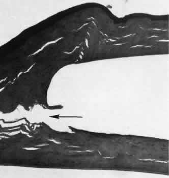
| Fig. 53. Light micrograph of an intracorneal pocket, which contained an intracorneal lens placed in an animal cornea. The contours of the pocket are smooth. There is no inflammatory reaction in the region of the lens. A artifactual crack of the corneal stroma in the posterior aspect of the wound (arrow) indicates the surgical plane of the incision used to implant the lens. (Periodic acid-Schiff stain; × 40.) |