|
|
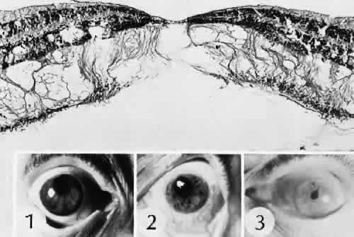
| Fig. 51. Cystoid macular edema following cataract extraction. In this case, no evidence of physical macular traction was identified. Microcystoid changes occur mainly in the outer retinal layers in this section though the fovea. (Hematoxylin-eosin stain; × 40.) Insets 1 to 3 show cases of pupillary distortion suggesting complicated cataract extraction and an increased risk for cystoid macular edema. |