|
|
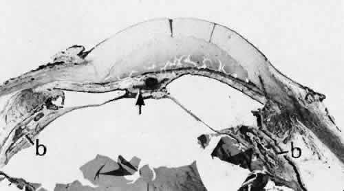
| Fig. 43. A case of extensive reactive uveitis following complicated ocular surgery. Inflammation generally causes increased adhesion of normally nonadhesive surfaces. In this case, both anterior synechiae and posterior synechiae have formed. Because the lens has been removed, the posterior synechia is between the iris and the vitreous face (arrow). The section is adjacent to the pupil. A small amount of iris sphincter muscle is in the region of posterior synechia formation. The anterior chamber is filled with a fluid containing a high concentration of protein, the result of prolonged inflammation. The abnormal biochemical environment has stimulated the formation of bone (b) in the anterior uveal tract. Dystrophic calcification and osseous metaplasia are common findings in degenerating eyes. Both types of deposits may be seen clearly by radiographic imaging. In children, this finding may be mistaken for calcification associated with retinoblastoma. (Hematoxylin-eosin stain; × 6.) |