|
|
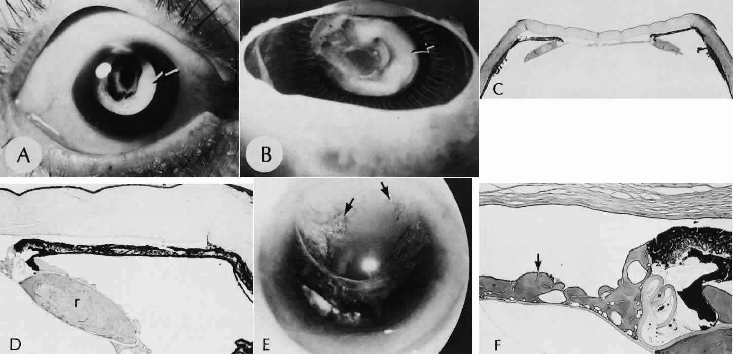
| Fig. 41. Two cases of Soemmerring's ring cataract (retained lens cortex and capsule). A. Extensive opaque, well-delineated material is present in a ring-shaped configuration following a partially successful cataract extraction. The opacity is delimited by the remaining lens capsule and is located primarily in the region of the lens equator. B. The eye was examined at autopsy. The reaction is limited to the posterior chamber and within the lens capsule. Surrounding lens zonules and ciliary epithelium are not affected. C. Lens cortical material is retained in the equatorial area of the lens. This area is the least surgically accessible during cataract extraction and contains the tissue most likely to be left behind. This area also contains the cells with the greatest ability to react to trauma by undergoing fibrous metaplasia. The process is identical to anterior subcapsular cataract formation stimulated by anterior chamber inflammation. (Hematoxylin-eosin stain; × 6.) D. Residual lens cortical material ® can be clearly identified entrapped by residual lens capsule. The reaction in the lens may change the adhesive nature of the remaining anterior lens capsule. Posterior anterior synechia may form in these areas. (Hematoxylin-eosin stain; × 16.) E. In another case of retained lens material, the tissue change is less extensive and more translucent, resulting in pearl-like structures (Elschnig's pearls). The process producing this change is the same as the one in Soemmerring's ring formation, but the reaction is less extensive. Pearls are formed by aberrant attempts by lens cells to form new lens fibers. (Hematoxylin-eosin stain; × 69.) |