
|
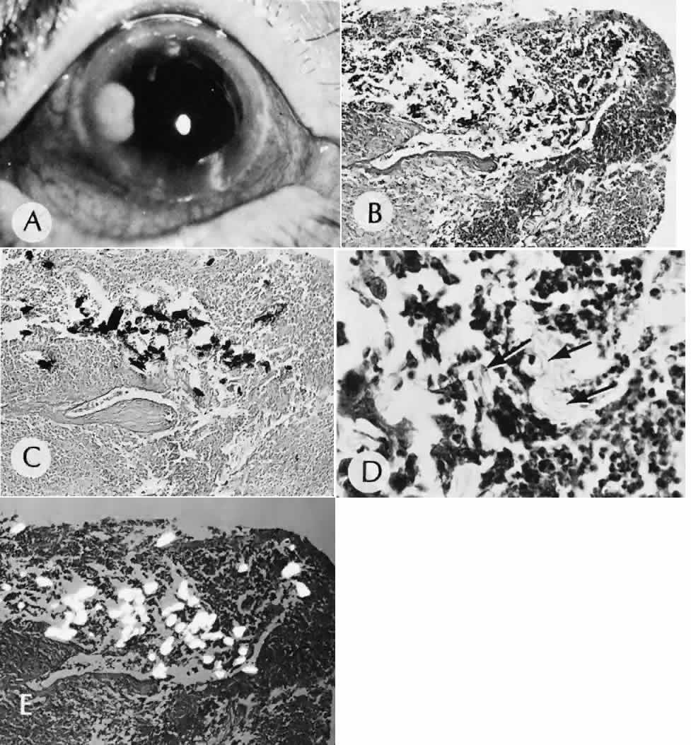
| Fig. 40. A case of suspected fungal endophthalmitis following cataract extraction. A. Well-demarcated, globular, opaque masses have developed over a period of weeks in the anterior chamber of a patient who had undergone cataract extraction. The lack of acute inflammatory activity, such as hypopyon formation, suggests a fungal infection. B. The anterior chamber reaction consists of a chronic inflammatory infiltrate characterized by multiple epithelioid histocytes, that is, a granulomatous inflammatory reaction consistent with fungal infection. (Hematoxylin-eosin stain; × 75.) C. A stain for fungus, however, does not reveal the presence of fungal forms. The material does stain with the silver stain, but the morphology is not that of a fungus. The features of the material suggest the inclusion of foreign material. (Gomori methenamine silver stain; × 75.) D. The material in the granulomatous inflammatory infiltrate appears to have a refractile nature when viewed at high magnification. (Hematoxylin-eosin stain; × 300.) E. The definitive test for foreign material is examination by polarized light. Under these conditions, the material can be identified as fibrous material consistent with cotton fibers. Cotton was apparently inadvertently introduced at the time of surgery. The inflammatory reaction is attempting to rid the eye of this foreign material, but the reaction is simultaneously destroying delicate ocular tissue. (Polarized hematoxylin-eosin stain; × 75.) |