|
|
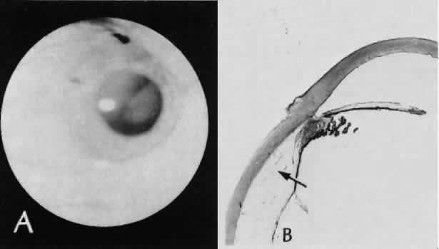
| Fig. 31. A case of choroidal detachment. A. By fundus reflex, a large dome-shaped mass can be seen originating from the choroid. The differential diagnosis would include uveal malignant melanoma. In this case, the clinical findings were due to choroidal detachment from the sclera because of fluid accumulating in the suprachoroidal space following cataract surgery. B. The histologic section from another case of choroidal detachment illustrates the location (arrow) and extent of the detachment. In this case the detachment extends to the region of the ciliary body limited anteriorly by the attachment of the choroid to the scleral spur. The displacement of the ciliary body will result in apparent shallowing of the anterior chamber. (Hematoxylin-eosin stain; × 6.) |