|
|
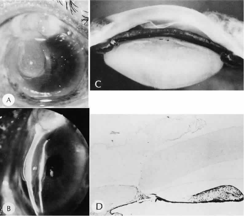
| Fig. 27. A case of stripping of Desce-met's membrane during a filteringprocedure (scleral cautery and iridectomy). A. The clinical appearance was one of extensive filtering bleb formation superiorly and regional dense corneal edema and opacification. B. The anterior chamber remains formed. Detached Descemet's membrane can be seen protruding into the anterior chamber. The patient died shortly after surgery from unrelated causes.C. The gross appearance of the area of detached Descemet's membrane extending into the anterior chamber.D. On the histologic section, the origin of detached Descemet's membraneextends to the region of the limbal wound. (Hematoxylin-eosin stain; × 16.) (Kozart DM, Eagle RC Jr: Stripping of Descemet's membrane after glaucoma surgery. Ophthalmic Surg 12:420–423, 1981.) |