|
|
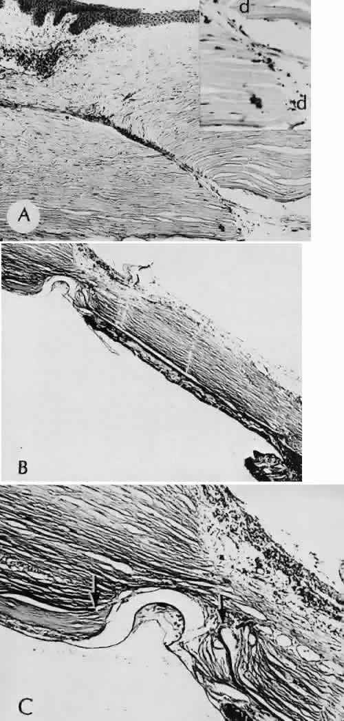
| Fig. 26. Complications related to a limbal wound. A. The posterior edges of the limbal wound are poorly apposed. Incarcerated vitreous can be identified in the wound at higher magnification, (inset). The cut edges of Descemet's membrane are widely displaced (d). (Hematoxylin-eosin stain; A, × 54; inset, × 101.) B. Vitreous is incarcerated into the wound immediately anterior to an area of total anterior synechiae. C. At higher magnification, vitreous can be clearly identified in the wound. A fibrous membrane is present posterior to Descemet's membrane. The arrows indicate the cut edges of Descemet's membrane. (Periodic acid/Schiff stain; B, × 16; C, × 40.) |