|
|
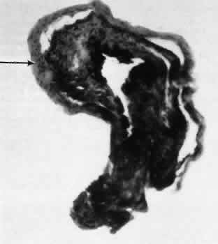
| Fig. 22. Light micrograph of an iridectomy specimen removed during a trabeculectomy procedure for secondary open-angle glaucoma following cataract extraction. The central iris tissue is covered with stratified squamous epithelial cells (arrow), indicating that epithelial ingrowth is present in this case. (Periodic acid-Schiff stain; × 100.) |