|
|
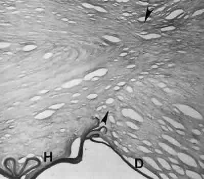
| Fig. 18. Light micrograph of a healed penetrating keratoplasty incision. The healed collagenous portion (between arrowheads) is barely visible. The host Descemet's membrane (H) is considerably thicker (older) and abnormal relative to the donor Descemet's membrane (D). A few redundant areas of Descemet's membrane are present at the graft-host interface. (Periodic acid-Schiff stain; × 40.) |