|
|
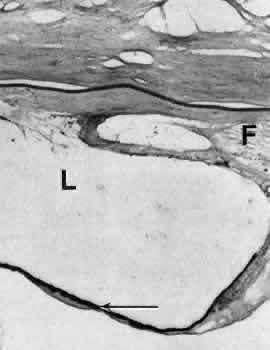
| Fig. 17. Light micrograph of fibrous tissue (F) completely filling the anterior chamber in a complicated case of pseudophakia. The outline of the lens optic (L) lies within the proliferated fibrous tissue and anterior to the remnants of the iris pigment epithelium (arrow). |