|
|
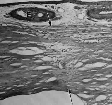
| Fig. 14. Light micrograph of a well-healed limbal wound. The wound through the sclera has healed almost completely, marked only by several small caliber blood vessels. Small epithelial inclusions (E) mark the site of the overlying conjunctival incision. Descemet's membrane in this specimen remains relatively straight. (Periodic acid-Schiff stain; × 40.) |