|
|
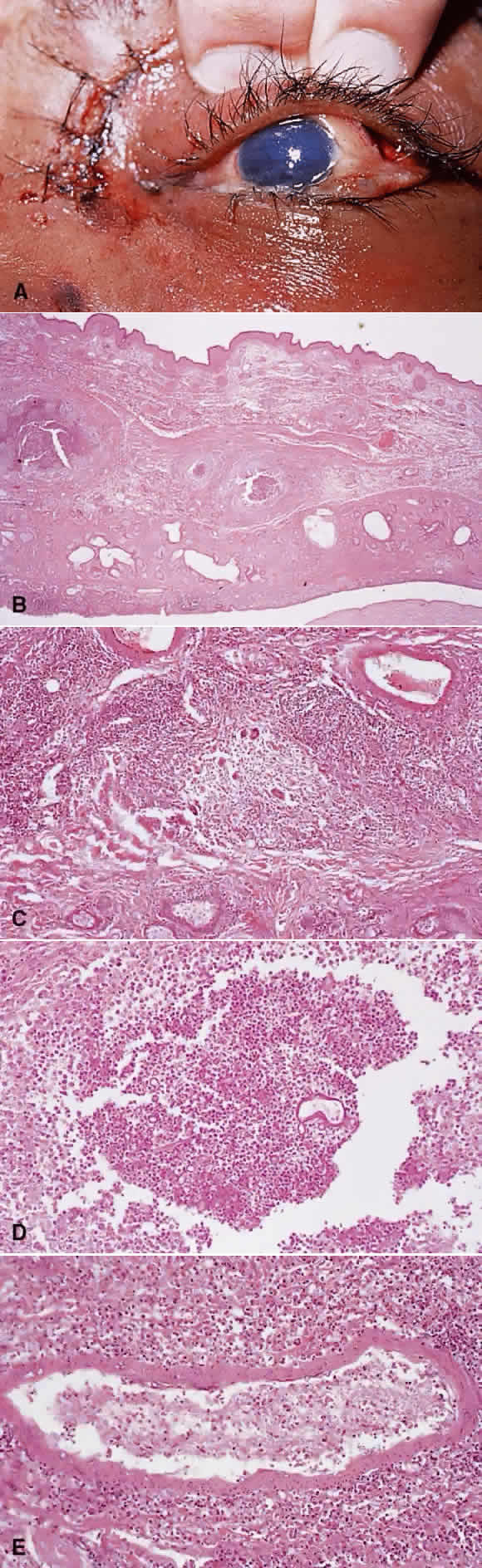
| Fig. 10. Mucormycosis. A. Young diabetic man with mucormycosis. Sutures are from biopsy. There is necrosis of the globe and orbit due to thrombosis of blood vessels. B. Full-thickness lid tissue with multiple granulomas in mucormycosis. C. Many giant cells within the granuloma. D. PAS-positive Mucor organisms, several in cross-section and one showing the broad, branching nonseptate hyphae. E. A posterior ciliary artery with many hyphae within the wall and lumen of blood vessel. |