|
|
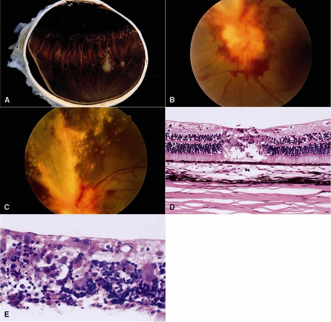
| Fig. 5. CMV retinitis. A. The gross specimen showing granular white patch of CMV retinitis at the ora serrata in an immunosuppressed renal transplant patient. B. Optic nerve swelling with retinal inflammation and hemorrhage. C. Sectoral area of atrophy surrounded by active CMV retinitis and hemorrhage. D. The normal retina suddenly becomes necrotic in the area of viral infection. E. High magnification of the retina with characteristic “owl's eye” intranuclear inclusions. |