|
|
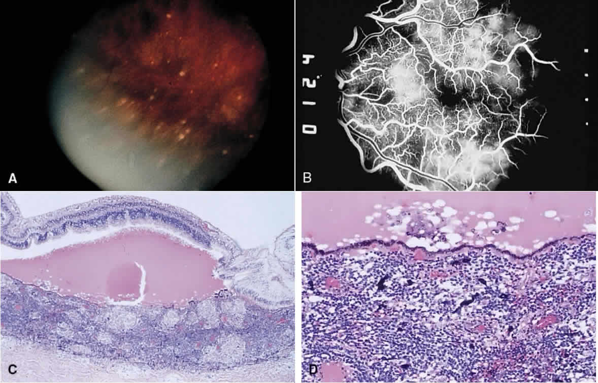
| Fig. 2. Sympathetic ophthalmia. A. Subretinal deposits in the midperipheral retina corresponding to Dalen-Fuchs nodules seen histopathologically. B. Fluorescein angiogram of multifocal areas of leakage at the level of the RPE. (A and B courtesy of William C. Frayer, MD.) C. Exudative retinal detachment and diffuse granulomatous inflammation in the choroid. D. The inflammation consists primarily of epithelioid cells and small lymphocytes. The choriocapillaris is spared. |