|
|
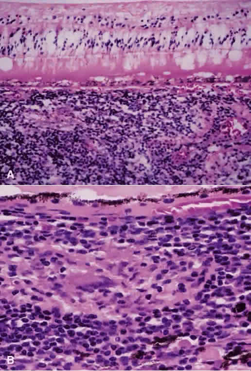
| Fig. 25. A. Retina and choroid of eye with sympathetic ophthalmia showing marked thickening of the choroid due to infiltration with chronic inflammatory cells (hematoxylin and eosin; × 200). B. Granuloma formation in the choroid. There is a loss of melanocytes in this region (hematoxylin and eosin; × 500). |