|
|
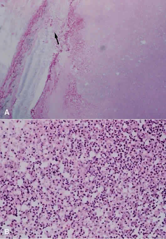
| Fig. 15. A. Photomicrograph of an eye enucleated for acute purulent bacterial endophthalmitis. Cells at right of figure are polymorphonuclear leukocytes with admixed hemorrhage. There is destruction of the retina (arrow) (hematoxylin and eosin; × 31). B. Higher power showing purulent material composed of viable and degenerating polymorphonuclear leukocytes and exudative material (hematoxylin and eosin; × 500). |