|
|
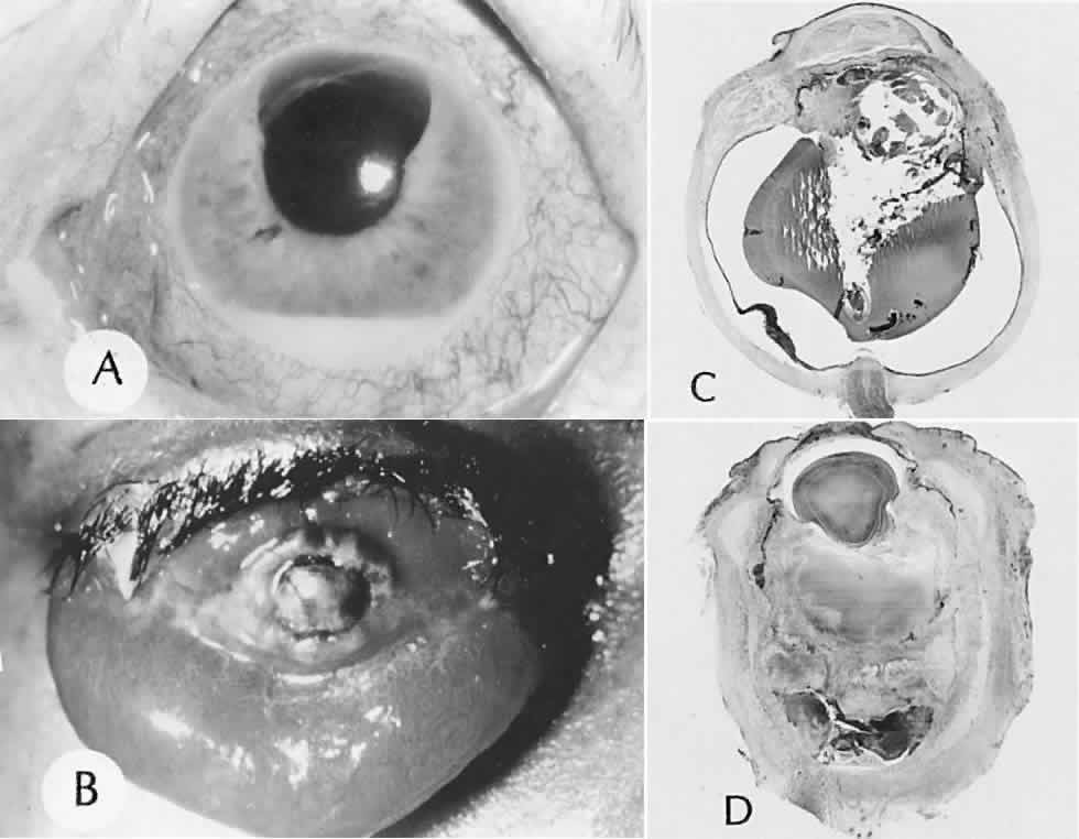
| Fig. 14. Endophthalmitis and panophthalmitis. A. Endophthalmitis developed in the patient following surgery for retinal detachment. Hypopyon is seen. B. Panophthalmitis following perforating surgery. Massive chemosis can be seen. C. Endophthalmitis shown by a vitreous adscess and iridocyclitis. The sclera contains no inflammatory reaction. D. Panophthalmitis shown by involvement of the vitreous cavity and all coats of the eye including the sclera. The cornea has perforated. (A and B, clinical [SEI 79-5 and 79-6]; C and D, hematoxylin and eosin, × 3 [SEI 79-7 and 79-8]. |